
After a stroke, two of the most important questions are: what caused it, and how can the risk of another stroke be reduced? When the cause of an ischaemic stroke remains unknown despite appropriate investigations, it is called a cryptogenic stroke. In selected patients, further assessment includes looking for a passage that allows blood to flow from the right to the left side of the heart. A bubble test — an echocardiogram with agitated saline contrast — can help detect this passage.
What Is the Connection Between the Heart and Stroke?
A patent foramen ovale (PFO) is a persistent flap-like opening between the right and left atria, the two upper chambers of the heart. This opening is normally present before birth and usually closes afterwards. However, it remains open in approximately one in four adults. Most people with a PFO have no symptoms and never experience a stroke.
Under certain circumstances, a PFO can allow a blood clot from the veins to cross into the arterial circulation, travel to the brain and block a blood vessel. This mechanism is called paradoxical embolism. For this reason, investigating a possible PFO can be an important part of the assessment of selected patients with a stroke of unknown cause.
How Is a Bubble Test Performed?
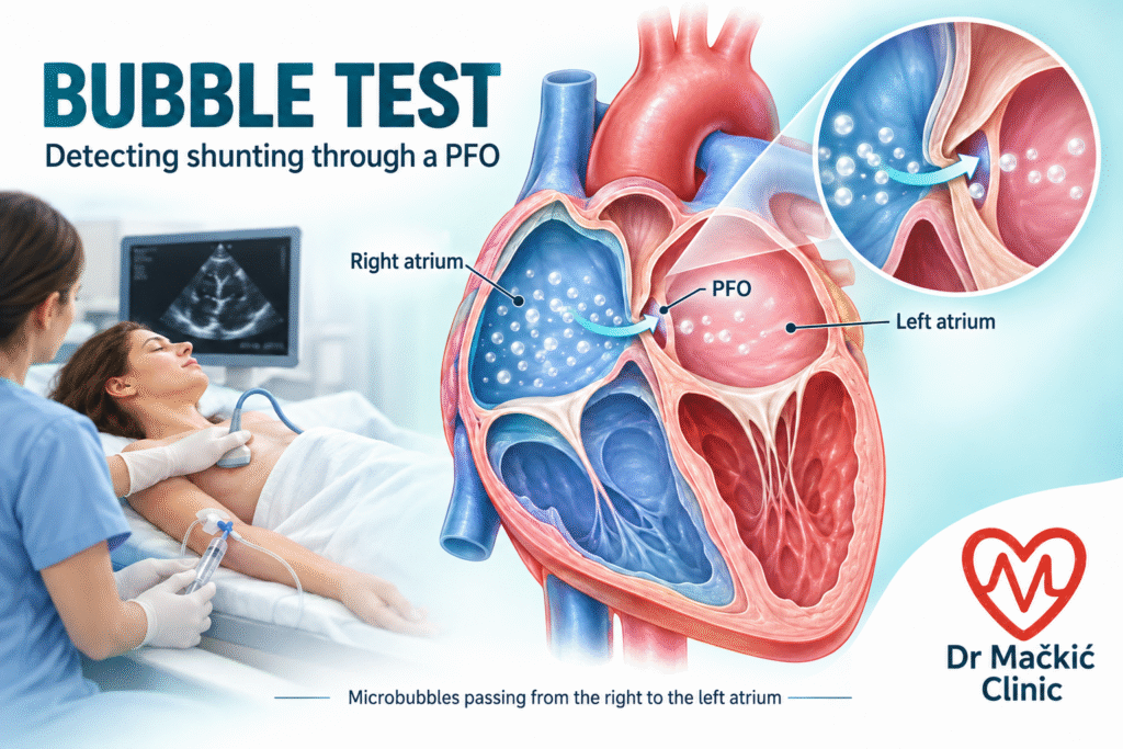
The examination uses an ultrasound probe placed on the chest. A specially prepared agitated solution containing tiny bubbles is injected through a cannula in a vein, while their movement is observed on ultrasound.
The bubbles first fill the right heart chambers. Their appearance in the left heart may indicate a right-to-left shunt, a passage that requires further assessment. The timing and pattern of bubble appearance help distinguish a passage through the heart from one through blood vessels in the lungs.
During the examination, the patient may be asked to briefly strain, as if having a bowel movement, and then relax. This is called the Valsalva manoeuvre. It can help reveal a PFO that does not allow bubbles to cross at rest. This examination through the chest does not require a probe in the oesophagus and does not use ionising radiation.
What Does a Positive Result Mean?
A positive bubble test does not automatically mean that a PFO caused the stroke or that it needs to be closed. The result is assessed alongside brain imaging, blood vessel investigations and heart rhythm monitoring, particularly to look for atrial fibrillation. When necessary, transoesophageal echocardiography is performed to examine the anatomy in greater detail.
In carefully selected patients, particularly those aged 18–60 years, PFO closure together with appropriate medical treatment can reduce the risk of another stroke. The decision is made jointly by a neurologist and a cardiologist following a comprehensive assessment.
Bubble Testing at Dr Mačkić Clinic
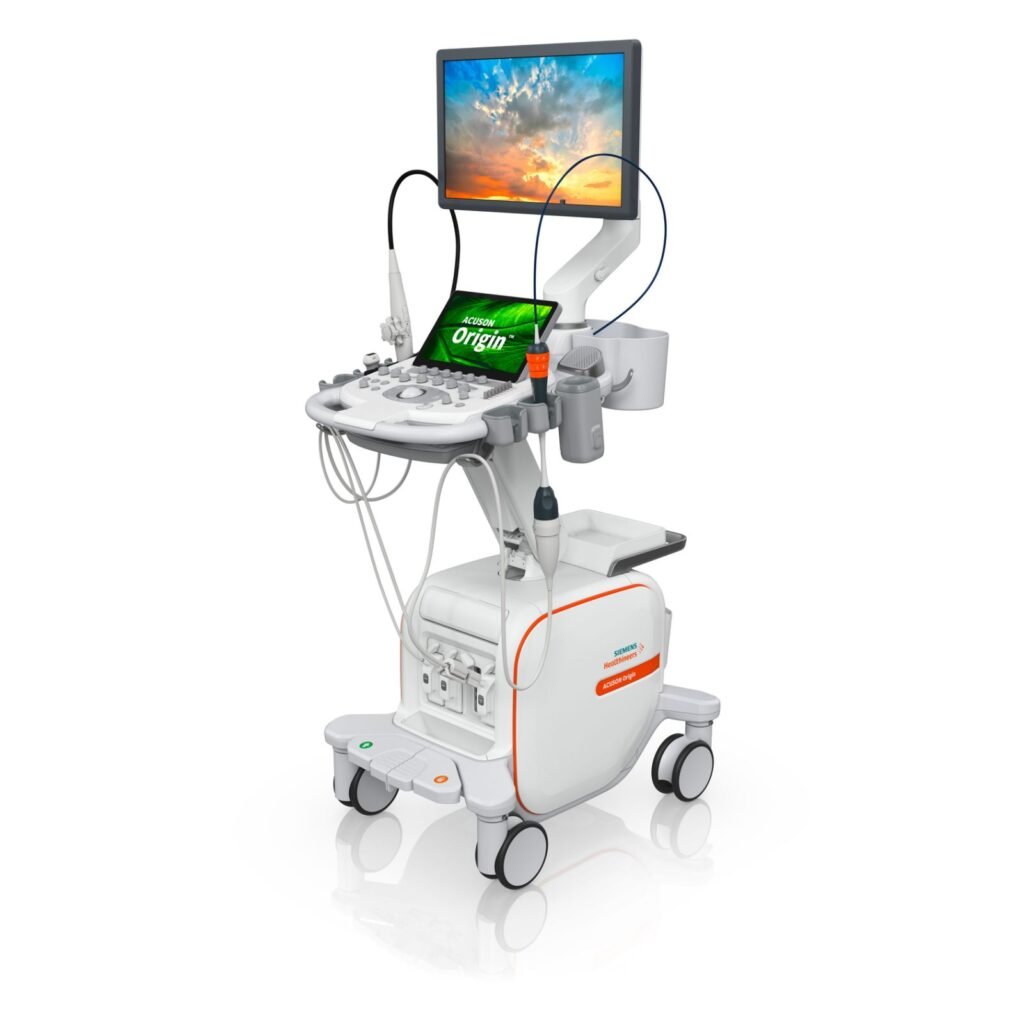
At Dr Mačkić Clinic in Sarajevo, bubble testing is available using the advanced, next-generation Siemens ACUSON Origin ultrasound system, which offers sophisticated cardiovascular imaging capabilities and technologies supported by artificial intelligence. Test is preformed by cardiologist Milan Gluhović MD, MA, FESC, FHFA.
The examination includes a cardiology assessment, performance of the bubble test and interpretation of the findings, with recommendations for further investigations when needed.
If you have experienced an ischaemic stroke whose cause remains unexplained, speak with your neurologist or cardiologist about whether a bubble test is an appropriate next step. To book an appointment, contact Dr Mačkić Clinic and bring your previous neurological and cardiology reports.
References
- Rana B, Robinson S, Ring L, Oxborough D, Belham M, Newton J, et al. Agitated saline contrast echocardiography in the diagnosis of right to left shunts: guidance and recommendations from the British Society of Echocardiography. Echo Res Pract. 2026;13:16. doi:10.1186/s44156-026-00117-3.
- Caso V, Turc G, Abdul-Rahim AH, Castro P, Hussain S, Lal A, et al. European Stroke Organisation (ESO) Guidelines on the diagnosis and management of patent foramen ovale (PFO) after stroke. Eur Stroke J. 2024;9(4):800-34. doi:10.1177/23969873241247978.
- Silvestry FE, Cohen MS, Armsby LB, Burkule NJ, Fleishman CE, Hijazi ZM, et al. Guidelines for the echocardiographic assessment of atrial septal defect and patent foramen ovale: from the American Society of Echocardiography and Society for Cardiac Angiography and Interventions. J Am Soc Echocardiogr. 2015;28(8):910-58. doi:10.1016/j.echo.2015.05.015.
- Siemens Healthineers. ACUSON Origin cardiovascular ultrasound system [Internet]. [cited 2026 Oct 10]. Available from: https://www.siemens-healthineers.com/ba/ultrasound/cardiovascular/acuson-origin.